Microslides, Animal Meiosis
Item code
2002092
Microslides, Animal Meiosis
$20.50
$22.55
Call us 1300 330 232
Microslides, Animal Meiosis
Microslides, Animal Meiosis
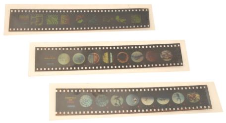
Microslides are sets of 8 related 35mm images photographed through a microscope called photomicrographs. Arrows and call outs help the student locate important features. The film is mounted in a clear plastic holder that protects it on both sides. Each Microslide is accompanied by a detailed lesson plan designed to stimulate, inform and question students about the topic under study. To be viewed using a Microslide Viewer (see item code 1002001).
This set contains images of:
the primary oocyte (tetrads formed, dyads separated, tetrads on spindle, first polar body forming), the secondary oocyte (chromosomes formed), the mature egg, the pronucleus stage, and the zygote ready for first cleavage.